
Why choose a Blood Component Separator when whole blood contains several valuable therapeutic elements? A validated separator can divide red cells, plasma, and platelets with greater consistency. Each component serves different clinical needs. This separation helps laboratories prepare targeted products instead of using unnecessary blood volume.
In a busy blood center, the difference appears in practical details. Staff can process labeled collection bags, monitor pressure, and inspect component layers during centrifugation. Modern systems may support controlled separation, sealed processing, and traceable records. These features can reduce handling steps and support safer, more efficient workflows. However, performance depends on trained operators, suitable equipment, and carefully verified procedures. Technology is helpful. It is not a substitute for judgment.
Experienced laboratory professionals also consider throughput, maintenance, compatibility, training, and service support. A separator should fit the facility’s collection methods and quality system. Independent validation remains important before routine use. Manufacturers may describe impressive specifications, but real performance can vary with bag design, blood properties, and operating conditions. That point is easy to overlook.
The best choice is therefore not always the fastest machine. It is the system that delivers repeatable results under local conditions. Reliable documentation matters. So does responsive technical support. When these factors align, a Blood Component Separator can strengthen component quality, improve resource use, and support more focused transfusion care. Yet every facility should review its own evidence carefully. No device solves every workflow problem.
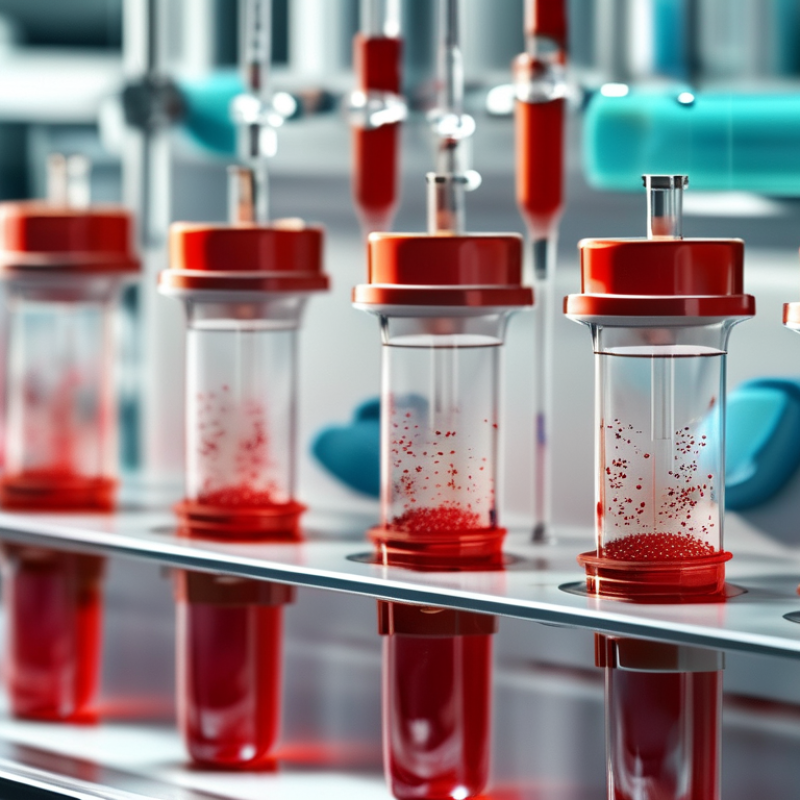
A blood component separator is a clinical device that divides whole blood into useful components. It commonly uses controlled centrifugation to separate red cells, plasma, and platelets by density. Some systems use apheresis to collect one selected component during donation or treatment. Different jobs.
Red cells carry oxygen, plasma contains important proteins, and platelets support normal clotting. Separating them allows each component to be stored and used according to its clinical purpose. In practice, staff load collection bags, select validated settings, and monitor the process carefully. A cloudy plasma bag, unusual color, or weak separation may require investigation. Small details matter. Temperature control, gentle handling, accurate labeling, and complete traceability help protect product quality and patient safety.
Choosing a blood component separator requires more than checking speed or capacity. The equipment should fit the facility’s workflow, staff training, maintenance resources, and applicable medical regulations. Reliable systems need calibration records, clear alarms, and procedures that reduce contamination risks. Operators also need practical training, not only a quick demonstration. Even a well-designed separator can be misunderstood. That is the uncomfortable part. A fast cycle is not automatically a better one. Reviewing errors and near misses can reveal whether the device truly supports consistent work. Clear documentation then helps teams adjust procedures before a small problem becomes a serious one.

A blood component separator works by exploiting differences in density. After collection, a centrifuge spins whole blood at controlled speed. Red cells move outward, while lighter plasma remains closer to the center. Between them, the buffy coat contains platelets and white blood cells. The device then transfers each layer into separate containers through sterile tubing.
The process is carefully timed. Sensors monitor pressure, volume, and fluid levels during separation. In automated systems, pumps guide selected components into collection bags while returning unwanted cells to the donor. This approach can collect more platelets from one session than a single whole-blood donation. However, performance depends on donor condition, anticoagulant control, and precise calibration. Small errors matter.
The World Health Organization’s Global Status Report on Blood Safety and Availability 2021 recorded 118.5 million blood donations worldwide in 2018. About 40% came from high-income countries, although those countries represented only 16% of the global population. These figures highlight why efficient component processing matters. One donation may support different clinical needs, including red-cell replacement, plasma therapy, or platelet treatment. The same report also emphasizes quality systems, trained staff, and reliable equipment. In practice, separation is not simply a spinning step. It requires validated settings, balanced rotors, temperature control, and documented checks. Some workflows still rely heavily on manual judgment, which deserves continued review.
| Data Dimension | How It Works | Typical Output or Result | Practical Benefit |
|---|---|---|---|
| Primary separation principle | Whole blood is separated mainly by centrifugation, which distributes components according to density. | Plasma forms the upper layer, the buffy coat contains most white blood cells and platelets, and red blood cells form the lower layer. | Supports consistent processing of donated blood into components with different clinical uses. |
| Component extraction | After centrifugation, controlled pressure or a press mechanism moves selected layers through sterile tubing into satellite bags. | The process can produce red-cell, plasma, platelet-rich, or cryoprecipitate-related components, depending on the collection and processing method. | Allows one whole-blood donation to be processed for use in different patients and clinical situations. |
| Red blood cell component | Red cells are separated from plasma and the buffy coat, then stored in an approved additive solution when applicable. | A concentrated red-cell component used to improve oxygen-carrying capacity. | Provides targeted red-cell support while reducing unnecessary exposure to other blood components. |
| Plasma component | Plasma is expressed from the separated upper layer and frozen or stored according to applicable blood-bank procedures. | A fluid component containing water, electrolytes, albumin, immunoglobulins, and clotting factors. | Enables component-specific replacement of plasma and selected coagulation factors. |
| Platelet preparation | Platelets are collected from platelet-rich plasma or from the buffy-coat layer through additional processing steps. | A platelet component that contributes to primary hemostasis and platelet plug formation. | Helps blood services prepare platelet products for patients with clinically significant thrombocytopenia or platelet dysfunction. |
| Leukocyte reduction | A validated leukocyte-reduction filter or processing step removes most white blood cells from a component. | A red-cell or platelet component with a substantially reduced leukocyte content. | May reduce febrile non-hemolytic transfusion reactions and exposure to donor leukocyte antigens when clinically indicated. |
| Closed-system processing | Sterile tubing, sealed collection bags, and validated connectors help maintain a closed fluid path. | Reduced need to expose the blood product to the surrounding environment during processing. | Supports aseptic handling and helps preserve component quality when procedures are followed correctly. |
| Standardization | Controlled pressure, calibrated scales, programmed separation steps, and documented operating procedures help regulate extraction. | More reproducible component volumes and fewer manual handling variations than an entirely manual workflow. | Improves process consistency, traceability, and staff workflow in blood-processing laboratories. |
| Quality control points | Operators verify identification, weights or volumes, seals, storage conditions, and applicable component quality specifications. | Traceable products that can be released only after required checks and testing are completed. | Helps protect product integrity and supports compliance with local blood-safety requirements. |
| Why choose a separator? | The device combines controlled layer separation, component transfer, weighing, and workflow monitoring in one processing system. | Multiple blood components can be prepared from a single donation using a structured process. | Can improve efficiency, reduce manual variability, support safer handling, and help blood centers use donated blood more effectively. |
Note: Exact component yields, processing settings, storage periods, and release criteria depend on the collection system, centrifugation protocol, equipment validation, and applicable national regulations.
A blood component separator turns one donation into targeted therapeutic products. It can isolate red blood cells, plasma, and the buffy coat, which contains most platelets and white blood cells. With controlled centrifugation, density becomes the working principle: red cells settle at the bottom, plasma stays above, and the buffy coat forms a narrow middle layer.
Apheresis systems separate components during donation. They can collect platelets, plasma, or red blood cells while returning other components to the donor. Plasma can also be frozen and processed into cryoprecipitate, a concentrated fraction containing fibrinogen, factor VIII, von Willebrand factor, and factor XIII.
White blood cells may be reduced through filtration, but that is different from isolating them as a final product. This distinction is often overlooked.
The need is substantial. The World Health Organization reported 118.5 million blood donations worldwide in 2021, with about 40% collected in high-income countries. Efficient separation helps hospitals use each donation more precisely, especially where supply is uneven.
However, separation is not automatically better. Poor temperature control, delayed processing, or incorrect centrifuge settings can damage cells and reduce product quality. In practice, trained staff, validated procedures, and continuous quality checks matter as much as the separator itself. The equipment helps, but it cannot replace judgment.
A blood component separator converts collected whole blood into red cells, plasma, and platelets. This supports targeted transfusion rather than giving unnecessary components. One donation may therefore serve several patients with different clinical needs. The World Health Organization’s Global Status Report on Blood Safety and Availability recorded 118.5 million blood donations worldwide in 2018. Efficient separation helps services manage this considerable resource more carefully.
Clinically, controlled separation can improve component consistency, reduce manual handling, and support clearer quality checks. Operationally, automated workflows may shorten processing time, strengthen traceability, and reduce avoidable waste. These benefits depend on trained staff, validated settings, and reliable maintenance. Equipment alone does not create safety. The SHOT Annual Report 2023 continues to highlight the importance of identification, documentation, and process discipline in transfusion practice. That is a useful warning. Faster processing can still fail when human checks become casual.
Tips: Match separator settings with validated component protocols. Monitor yield, temperature, and processing time for every batch. Review rejected units, not only successful ones. Keep staff training practical, with real samples and realistic interruptions. A simple dashboard can reveal recurring delays, but data may not explain every cause. Local workflow, donor characteristics, and storage capacity still matter. Teams should challenge impressive performance figures and confirm them against their own clinical results.
Why Choose a Blood Component Separator?
What Factors Guide the Selection of a Suitable Separator?
Selecting a blood component separator begins with workflow, not appearance. The device should match collection volume, processing speed, and staff capability. WHO reported 118.5 million blood donations across 169 countries in its 2021 global report. It also found that high-income countries collected 40% of donations, despite representing 16% of the global population. These differences make local workload and resource conditions essential selection factors.
Separation quality deserves close attention. Ask whether the system supports consistent plasma, platelet, and red-cell yields. Check hemolysis control, closed-system operation, traceability, and compatibility with existing collection bags. AABB quality standards emphasize validated processes, documentation, equipment maintenance, and staff training. These requirements are practical, not decorative. A separator with impressive throughput may still create delays if setup takes too long. That detail is easy to underestimate.
Power stability and service access also matter. A rural center may value low energy demand and simple maintenance more than maximum automation. The U.S. National Blood Collection and Utilization Survey has repeatedly shown major differences in collection capacity between facilities. Therefore, one specification cannot fit every laboratory. No separator is perfect. Selection teams should test real samples, review failure records, and compare usable output rather than brochure claims. I would also reassess the choice after several months, because operator feedback often reveals problems that controlled demonstrations miss.
Component type, processing speed, temperature control, closed-system operation, automation, traceability, and validated capacity are key factors when selecting a suitable separator.
The chart shows commonly used maximum storage durations for separated blood components: red blood cells up to 42 days at 1–6°C, platelets up to 7 days at 20–24°C when an approved bacterial-risk-control system is used, and plasma or cryoprecipitate up to 365 days when stored at −18°C or colder. Actual limits depend on local regulations, anticoagulants, processing methods, and validated equipment.
“Establish the work of our hands”
Psalm 90:17b